
Figure 8.
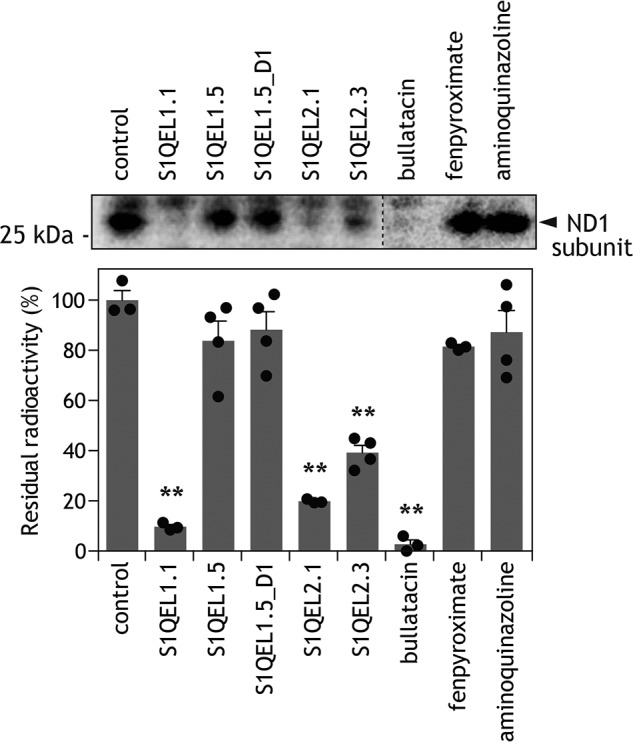
Effects of different inhibitors on the labeling of the ND1 subunit by [125I]S1QEL1.1_PD1. The photoaffinity labeling of the ND1 subunit by [125I]S1QEL1.1_PD1 (5.0 nm) was carried out in the presence of different inhibitors (5.0 μm each, 1000-fold of [125I]S1QEL1.1_PD1), as described under “Experimental procedures.” Proteins equivalent to ∼50 μg of SMPs were loaded into each well. Upper panel, gel image of SDS-PAGE analysis used for photoaffinity labeling; lower panel, the extent of suppression (values in graphs are mean ± S.E. (n = 3–4)). **, p < 0.001 compared with control (one-way ANOVA followed by Dunnet's test).