
Figure 9.
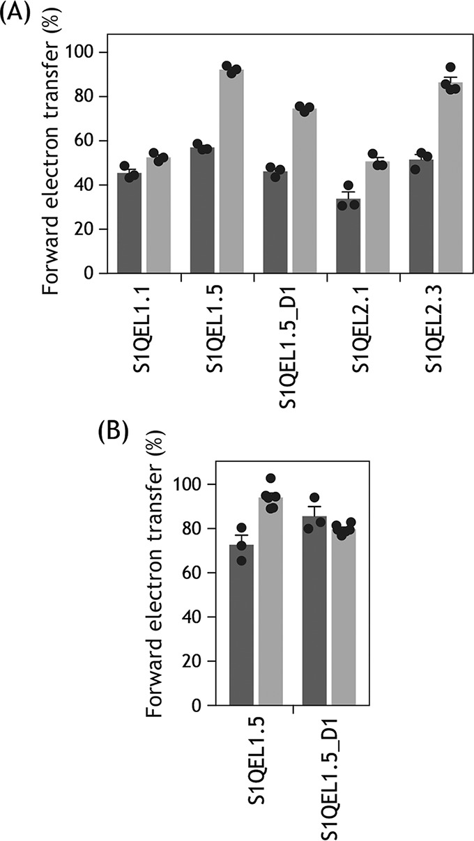
Effects of BSA on the inhibitory action of S1QELs. A, the inhibition of forward electron transfer by S1QELs was examined after incubation with SMPs for 4 min in the absence (black bars) or presence (gray bars) of 0.3% fatty acid-free BSA in assay buffer, except for S1QEL1.5 and S1QEL1.5_D1. The inhibition by S1QEL1.5 and S1QEL1.5_D1 was evaluated at ∼1 min after addition of the compound to respiring SMPs (cf. trace c in Fig. 2). The concentrations of S1QELs were: S1QEL1.1 (0.062 μm), S1QEL1.5 (1.2 μm), S1QEL1.5_D1 (2.0 μm), S1QEL2.1 (0.28 μm), and S1QEL2.3 (0.37 μm). B, the inhibition by S1QEL1.5 (1.2 μm) and S1QEL1.5_D1 (2.0 μm) was examined after incubation with SMPs for 4 min in the absence (black bars) or presence (gray bars) of 0.3% fatty acid-free BSA in assay buffer. Values in graphs are mean ± S.E. (n = 3–6).