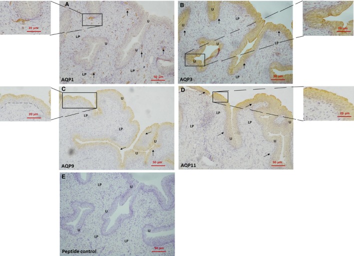
Figure 2.

Immunoperoxidase labelling of AQPs in pig bladder. A, bladder mucosa with AQP1 immunoreactivity in the lamina propria. The inset shows a small blood vessel immediately below the urothelium at a larger magnification. B, bladder mucosa with AQP3 immunoreactivity in the urothelium. The inset shows the urothelium at a larger magnification. C, bladder mucosa with AQP9 immunoreactivity in the urothelium. The inset shows the urothelium at a larger magnification. The dotted line demonstrates the boundary between the urothelium and the lamina propria. D, bladder mucosa with AQP11 immunoreactivity in the urothelium. The inset shows the urothelium at a larger magnification. E, A representative bladder mucosa with peptide control. U, urothelium; LP, lamina propria
