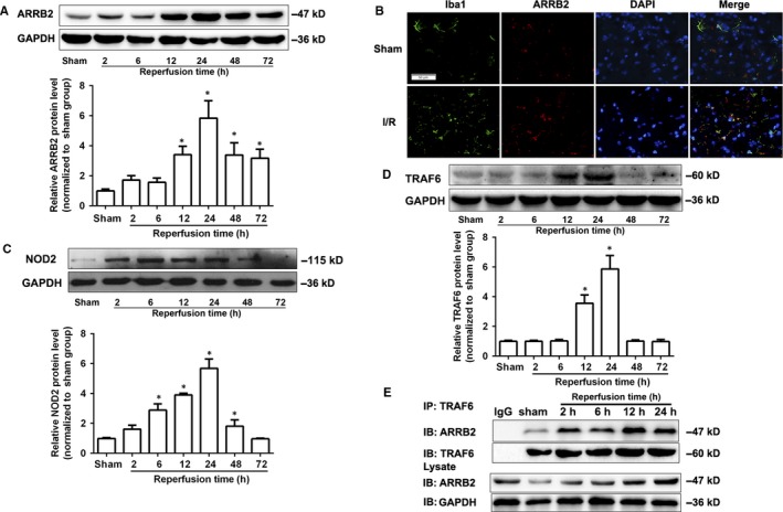
Figure 1.

The expression of β‐arrestin2 (ARRB2) in wild‐type (WT) mice was increased after cerebral ischaemia‐reperfusion (I/R) injury. Western blot analysis of ARRB2 (A), NOD2 (C) and TRAF6 (D) protein levels in the penumbral cortex from WT mice after 2 h occlusion of the middle cerebral artery (MCAO) and 2, 6, 12, 24, 48 and 72 h reperfusion. Results are representative of six independent experiments. *P < 0.05 compared with sham group. (B) Representative images of double immunolabelling for ARRB2 and Iba‐1(microglia) in the penumbral cortex from WT mice after 2 h MCAO and 24 h reperfusion. DAPI indicates 4′,6‐diamidino‐2‐phenylindole. Scale bars: 50 μm. (E) The interaction of ARRB2 and TRAF6 was determined by Co‐Immunoprecipitation (Co‐IP) in the penumbral cortex from WT mice after 2 h MCAO and 2, 6, 12 and 24 h reperfusion
