Figure 5.

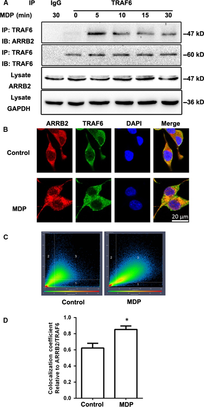
MDP stimulated interaction of β‐arrestin2 (ARRB2) with TRAF6. (A) BV2 cells were stimulated with MDP (2 μg/mL) for 5, 10, 15 and 30 min. Cell extracts were immunoprecipitated with anti‐TRAF6 antibody and then analysed together with cell lysate by immunoblotting with indicated primary antibodies. (B) BV2 cells were stimulated with MDP (2 μg/mL) for 5 min. ARRB2 (red) and TRAF6 (green) were viewed using confocal microscopy. Scale bars: 20 μm. (C) 2D fluorograms showing colocalization of ARRB2 and TRAF6 as a distribution of pairs of pixel intensities (with greater diagonal alignment correlating to higher colocalization). (D) Quantification of the colocalization coefficient between ARRB2 and TRAF6. *P < 0.05 compared with control group
