Figure 3.

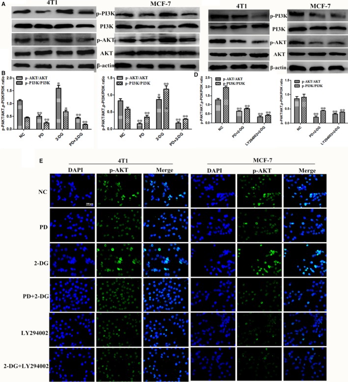
Polydatin (PD) combined with 2‐deoxy‐D‐glucose (2‐DG) inhibits the PI3K/AKT signalling pathway. The 4T1 and MCF‐7 cells were treated for 24 h with either 2‐DG alone or PD or LY294002 (2 μmol/L) or with a combination of both agents alone. (A‐D) The protein expression levels of p‐PI3K, PI3K, p‐AKT and AKT were detected by western blot. β‐actin was used as an internal control. (E) Immunofluorescence staining of p‐AKT in 4T1 and MCF‐7 cells treated with PD, 2‐DG and their combination. Scale bar: 100 μm (n = 3). All results are expressed as the mean ± SEM of three independent experiments. The symbols *and **denote significant differences of P < 0.05 and P < 0.01, respectively
