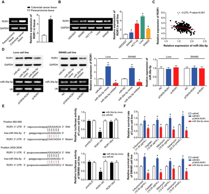
Figure 5.

The mediation of ROR1 for the contributions of XIST and miR‐30a‐5p to chemosensitivity of colorectal cancer cells. A, The expression of ROR1 was compared between colorectal cancer tissues and paracarcinoma tissues. *P < 0.05 when compared with paracarcinoma tissues. B, The expression of ROR1 was determined within HEK293T, SW480, HCT116, Lovo and SW620 cell lines. *P < 0.05 when compared with HEK293T. C, Among the incorporated colorectal cancer tissues, ROR1 expression was positively correlated with XIST expression, yet it displayed negative relevance to miR‐30a‐5p. D, The expression of ROR1 was detected after transfections of pcDNA‐XIST, s‐XIST, miR‐30a‐5p mimic and miR‐30a‐5p inhibitor, and the expressions of XIST and miR‐30a‐5p were also determined after transfections of pCMV1‐ROR1 and si‐ROR1. *P < 0.05 when compared with NC. E, ROR1 was subjected to target of miR‐30a‐5p in certain sites, and the luciferase activity of cells was compared among miR‐30a‐5p mimic+pmirGLO‐ROR1‐Wt, miR‐30a‐5p mimic+pmirGLO‐ROR1‐Mut and miR‐30a‐5p mimic+pmirGLO groups. *P < 0.05 when compared with pmirGLO+miR‐30a‐5p mimic group. F, The sensitivity of colorectal cancer cells was compared when responding to 5‐fluorouracil, mitomycin, cisplatin and adriamycin among the miRNA‐NC, miR‐30a‐5p mimic and miR‐NC+pCMV1‐ROR1 groups. *P < 0.05 when compared with NC
