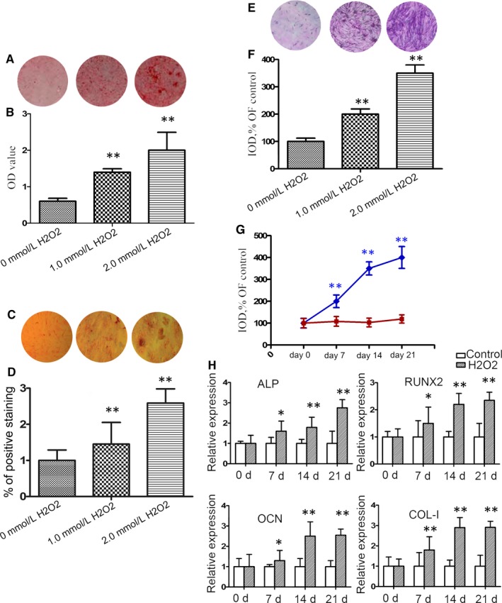
Figure 1.

Oxidative stress induces mineralization in primary endplate chondrocytes from rat intervertebral discs (IVDs). Endplate chondrocytes were isolated from rats and treated with, or without, H2O2 at the indicated doses for 7 days. (A) Alizarin Red staining for calcium deposition in endplate chondrocytes. (B) Semi‐quantitative analysis of the mineralized nodule in endplate chondrocytes. (C) von Kossa staining. (D) The percentage of von Kossa‐positive cells. (E) ALP staining in endplate chondrocytes. (F) Semi‐quantitative analysis of alkaline phosphatase (ALP) activities in endplate chondrocytes. (G) Longitudinal analysis of ALP activities in endplate chondrocytes following treatment with 2.0 mM H2O2. (H) Quantitative real time polymerase chain reaction (RT‐PCR) analysis of the relative levels of ALP, RUNX2, OCN and COL‐I mRNA transcripts in endplate chondrocytes. Data are representative images (magnification x 100) or expressed as the mean ± SEM of each group of cells from three separate experiments. *P < 0.05, **P < 0.01 vs the control
