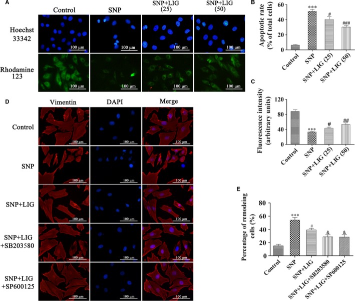
Figure 2.

Effects of ligustilide (LIG) on nuclear morphology, mitochondrial membrane potential and cytoskeletal remodelling in sodium nitroprusside (SNP)‐stimulated chondrocytes. (A, B, C) Hoechst 33342 and Rhodamine‐123 staining of chondrocytes exposed to LIG (25 and 50 μmol/L) for 2 h before 0.75 mmol/L SNP co‐treatment for 24 h. The levels of chondrocyte nucleic morphologic changes and intracellular Rhodamine‐123 fluorescence were evaluated. (D, E) Fluorescent images with Vimentin‐Tracker red of chondrocytes pre‐incubated with LIG (50 μmol/L) in the presence and absence of the JNK inhibitor SP600125 (10 μmol/L) and the p38 mitogen‐activated protein kinase (MAPK) inhibitor SB203580 (10 μmol/L) for 2 h before 0.75 mmol/L SNP co‐treatment for 24 h, and the cell nuclei were stained with DAPI. The percentage of remodelling cells was measured. Each column represented mean ± SEM (n = 5). ***P < 0.001 vs the control group; # P < 0.05, ## P < 0.01 and ### P < 0.001 vs the SNP group; & P < 0.01 vs the SNP + LIG (50 μmol/L) group
