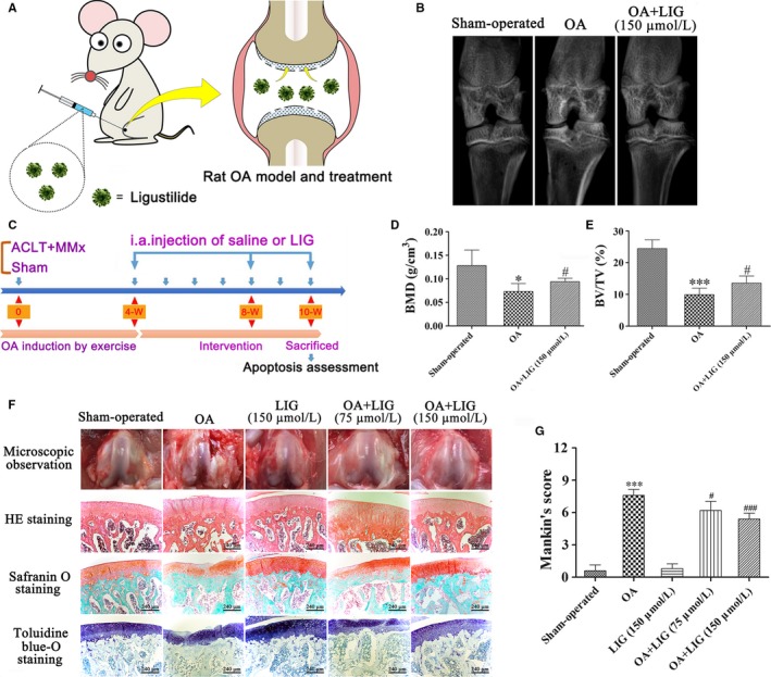
Figure 4.

Schematic depicting the time of treatment, the follow‐up period of the animals and the effects of ligustilide (LIG) on cartilage degradation in rat articular cartilage 10 weeks after anterior cruciate ligament transection together with medial menisci resection (ACLT + MMx). (A, C) ACLT + MMx rats were placed in an electric rotating cage for 30 min per day to induce the osteoarthritis (OA) model from the 1st week after surgery. Low‐ and high‐dose LIG treatment animals were injected intra‐articularly with 30 μL of 75 and 150 μmol/L LIG 4 weeks after surgery. The sham‐operated and OA‐induced animals received an injection of 30 μL of PBS. The protective effects of LIG on OA progression in vivo were assessed 10 weeks after surgery. (B) Representative micro‐computed tomography two‐dimensional reconstructions of tibial and femoral subchondral bones in the rats after ACLT + MMx surgery or sham operation. (D, E) Bone mineral density (BMD) and bone volume fraction (BV/TV) were measured in the subchondral bones of the knee joint of vehicle‐treated and LIG‐treated rats 10 weeks after ACLT + MMx surgery or sham operation. (F) Gross morphological and histological analyses of rat articular cartilage by H&E staining, Safranin O staining and toluidine blue‐O staining (original magnification 400 ×) in each group. (G) Overall Mankin's histological score was assessed in five groups 10 weeks after ACLT + MMx surgery or sham operation. Each column represented mean ± SEM (n = 5). *P < 0.05 and ***P < 0.001 vs the sham‐operated group; # P < 0.05 and ### P < 0.001 vs the OA‐induced group
