Figure 6.

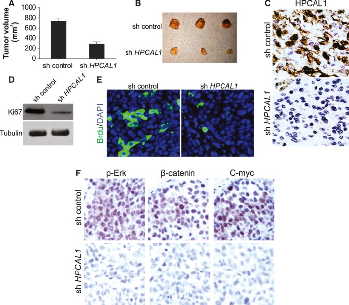
HPCAL1 enhances proliferation of GBM cell in vivo. (A) NOD‐SCID mice received subcutaneous injection of parent and HPCAL1 stably knockdown A172 cells to construct xenograft cancers. The volume of the malignancy was evaluated and recorded 3 weeks after the inoculation (n = 5/group). (B) Cancers at terminal stage in the experiment. (C) Representative immunochemistry staining of HPCAL1 in tumours from (B). (D) Western blotting (WB) of Ki67 in distinct groups of cancer from (B). (E) Representative immunostaining of Brdu in cancers from (B). (F) Immunochemistry staining analysis of p‐Erk, β‐catenin and c‐Myc in cancer specimens from (B). Western blots are representative pictures for two independent replicates
