
Figure 2.
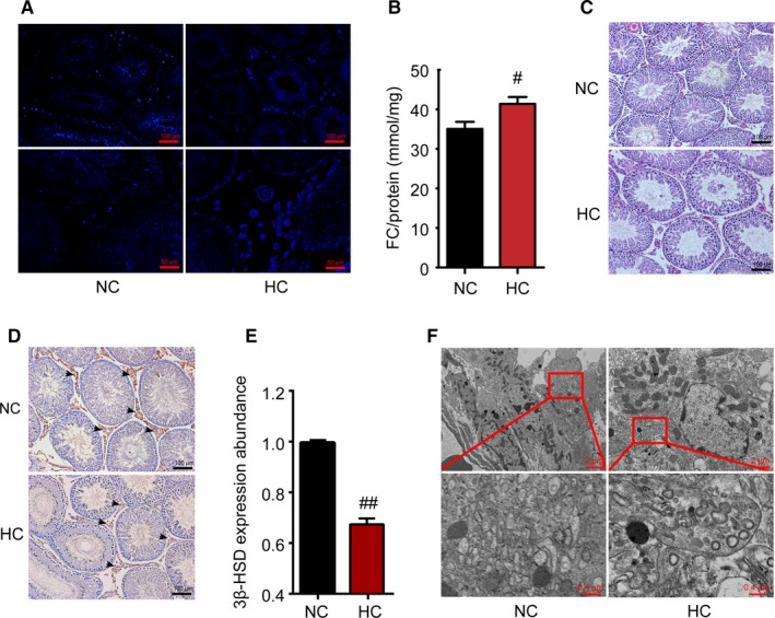
HC diet induces morphological changes in the testes. After HC‐diet feeding, filipin fluorescence was performed to display free cholesterol deposition in the testis gland in both groups (blue colour) (A). Further, testicular free cholesterol (FC) content was assayed and corrected by the total protein content (B). Representative photomicrographs of H&E staining of testicular sections are shown (C). D, Testis was immunostained for 3β‐HSD which is specifically expressed in Leydig cells, and 3β‐HSD expression abundance in the Leydig cells was analysed (E). The ultrastructure of the Leydig cells in the testis of the NC group revealed a normal fine structure. In the testes of the HC‐group rats, the dilated endoplasmic reticulum (red box) was observed in the Leydig cells (F). Data are presented as the mean ±SEM (n = 8 per group). #P < 0.05, ##P < 0.01 vs the NC group. All results were from at least three independent experiments