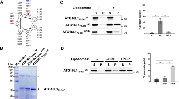
Figure 6. Negatively charged residues within the CCD of ATG16L1 weakens its interaction to lipids.

- ATG16L1 CCD region 164–198 depicted on a heptad repeat with the relevant residues highlighted in red (acidic; D164 and E165), purple (hydrophobic; I171) and blue (basic; K179 and R193).
- Coomassie gel of recombinant ATG16L172–307 wild type and mutants. * indicates bacterial protein.
- Liposome co‐sedimentation assay using recombinant ATG16L1 protein and sonicated liposomes. Supernatant (S) and pellet (P) were analysed by immunoblotting against T7 tag to detect ATG16L1. Quantification of percentage protein in the pellet fraction was calculated from three independent experiments (right panel). Error bars (SEM). **P ≤ 0.01 (unpaired Student's t‐test).
- Liposome co‐sedimentation assay, as in (C), using ATG16L172–307 LE and sonicated liposomes that contain or lack PI3P. Quantification of percentage protein in the pellet fraction was calculated from three independent experiments (right panel) with error bars depicting SEM values. ns P > 0.05, **P ≤ 0.01, ***P ≤ 0.001 (pairwise unpaired Student's t‐test).
