
Figure 2. Gata6 expression is independent of Wnt/β‐catenin signaling.
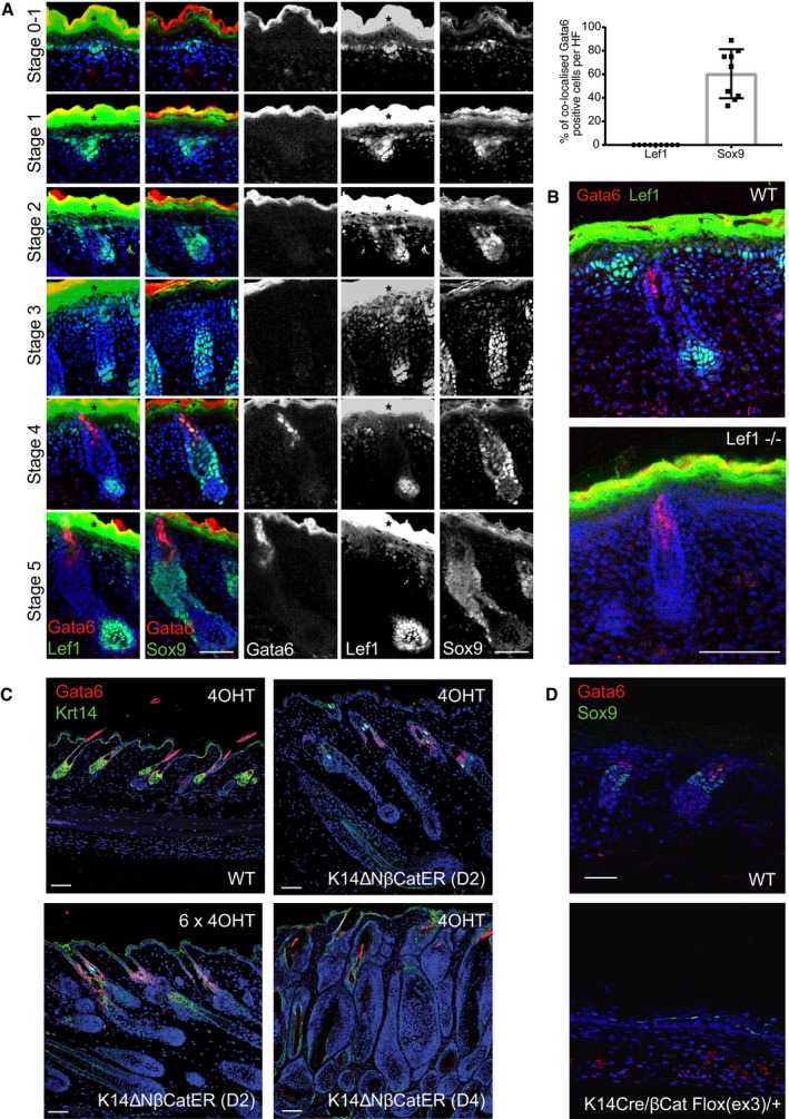
- Sections of WT embryonic skin at different HF stages stained for Gata6, Lef1 or Sox9, and counterstained with Dapi. Black asterisks indicate overexposed areas of nonspecific Lef1 staining in the suprabasal epidermis. Quantification of the percentage of cells labeled for both Gata6 and Lef1, or Gata6 and Sox9 in stage 4–5 HF is shown (upper right panel). Data are means ± SD and were obtained from 9 HF from 3 mice.
- Sections of E18.5 WT and Lef1−/− mouse skin stained for Lef1 and Gata6. Deletion of Lef1 does not impair Gata6 expression.
- Sections of WT and K14ΔNβ‐CateninER (K14ΔNβ‐CatER D2 and D4 strains) adult dorsal skin stained with antibodies against Krt14 and Gata6. Topical treatment with 1 or 6 doses of 4OHT activates β‐catenin, leading to anagen induction and ectopic hair follicles but not Gata6 expression.
- Sections of E18.5 WT and K14Cre/βCat Flox(ex3)/+ mouse skin stained for Sox9 and Gata6. Activation of Wnt/β‐catenin signaling during epidermis development does not induce Gata6 expression but results in ectopic expression of the HFSC marker Sox9.