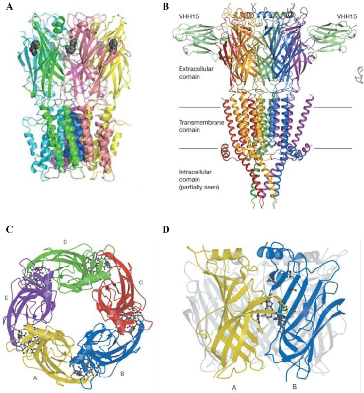
Figure 1.
(A) Structure of the α7-nAChR. A comparative model based on the homologue protein from Erwinia chrysanthemi (Protein Data Bank code 2VL0), side view. Figure adapted from Taly et al.24 (B) X-ray structure of mouse 5-HT3R in complex with the VHH15 stabilizing nanobody (Protein Data Bank code 4PIR, 3.50 Å resolution). Side view picture is shown. Figure adapted from Hassaine et al.214 (C,D) From x-ray structure of Lymnea stagnalis AChBP (Protein Data Bank code 1I9B, 2.7 Å resolution). (C) Top view, five subunits displayed. (D) Side view, displaying the ligand binding site between two subunits. Figures adapted from Brejc et al.215 License agreements for using these figures (A–D) were provided by the Copyright Clearance Center (CCC).

