
Figure 3.
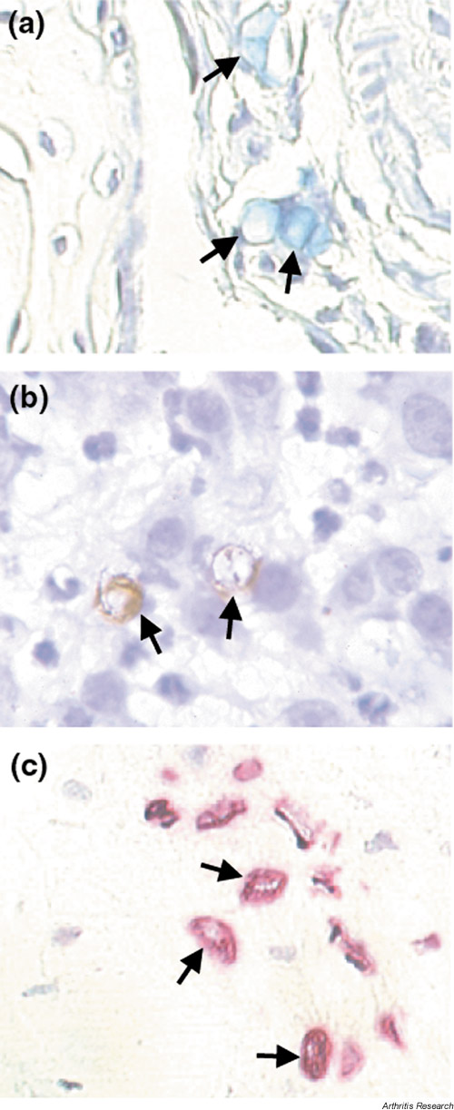
Detection of RGD-4C-phage, αvβ3, and TUNEL positive cells in synovial blood vessels. Mice with established collagen-induced arthritis were injected with RGD-4C and sections were stained for (a) phage (blue blood vessels, denoted here by arrows) or (b) –vβ3 (brown blood vessels, denoted here by arrows) by immunohistochemistry. In a second experiment, mice with collagen-induced arthritis were given RGD-4C-D(KLAKLAK)2 on day 35 and TUNEL assays were performed on day 38. Positive cells were found in sublining vessels of inflamed synovium in the animals treated with RGD-4C-D(KLAKLAK)2 (arrows) (c) but not in the control animals (not shown).