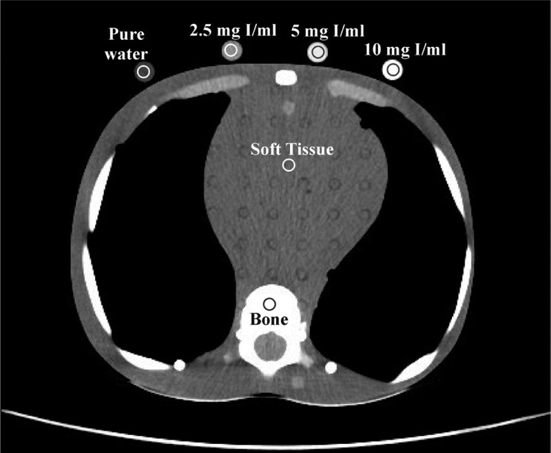
FIGURE 3.

An axial slice depicting thorax of the 10-year-old phantom. Shown are the ROIs drawn over soft tissue, bone, and iodinated and pure water vials at the axial plane depicting the middle heart level. All but bone ROIS were 70 mm2 in size. Bone ROIs were 20 mm2 in size. Protocol acquisition parameters for slice shown; clinical mode: CTA, 100 kVp, NI: 11.7, slice thickness 2.5 mm, CTDIvol 2.67 mGy.
