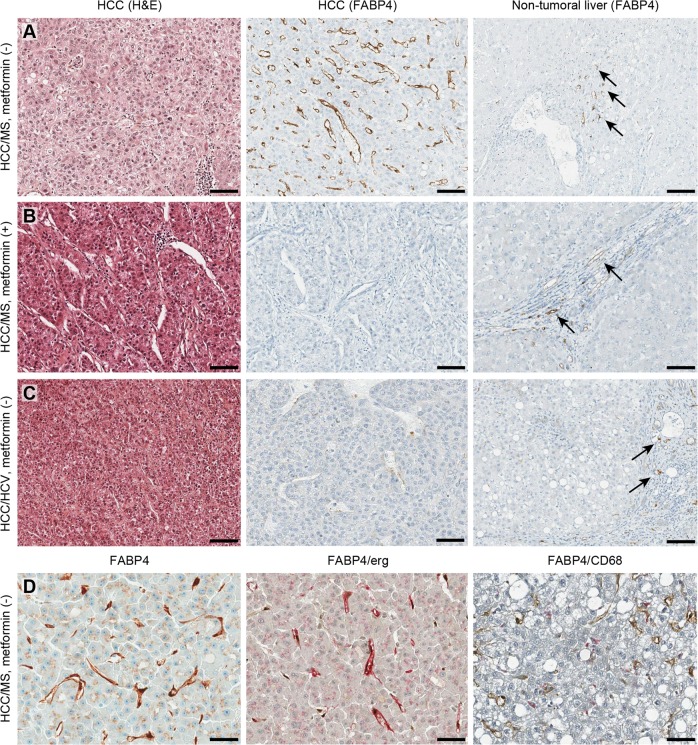
Fig. 2.
FABP4 is mostly expressed in endothelial peritumoral cells in HCC related to MS. a HCC/MS without metformin treatment. b HCC/MS with metformin treatment. c HCC/HCV. Three panels include hematoxylin & eosin staining of HCC (left), FABP4 immunostaining in HCC (middle) and FABP4 immunostaining in non-tumoral liver (right). Arrows indicate positive vessels in portal tracts and fibrous bands. (Scale bars = 100 μm). d FABP4 immunostaining in HCC/MS without metformin. (left) higher magnification showing slight staining was observed in tumoral hepatocytes; (middle) double immunostaining with FABP4 (pink) and ERG (endothelial cells, brown); (right) double immunostaining with FABP4 (brown) and CD68 (macrophages, pink)

