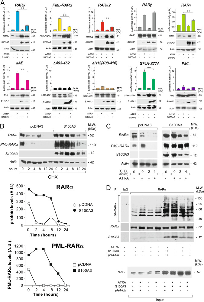
Fig. 3.
RARα structural determinants of the interaction with S100A3 and effects of S100A3 on the stability and ubiquitinylation of the retinoid-receptor and the PML-RARα fusion product. a COS-7 cells were transiently co-transfected with an expression plasmid for the S100A3 cDNA (S100A3) or the corresponding void vector (pcDNA3) and wild-type RARα, PML-RARα, RARα2, RARβ, RARγ, the indicated RARα deletion-mutants and point-mutants along with a luciferase reporter construct controlled by a retinoid responsive element (β2RARE-Luc). PML was used as an internal negative control of the experiment. Twenty-four hours following transfection, cells were treated with vehicle (DMSO) or ATRA (1 µM) for a further 24 h. Cell extracts were subjected to western blot analysis with anti-RARα, anti-RARβ, anti-RARγ, anti-PML (upper panels), anti-S100A3 (middle panels), and anti-actin (lower panels) antibodies. The same cell extracts were used for the measurement of luciferase activity, as illustrated by the bar graphs above the western blots. Each value is the mean ± SD of three replicate cultures. **Statistically significant comparison (p < 0.01, Student’s t-test). b COS-7 cells were transiently co-transfected with S100A3 or pcDNA3 and wild-type RARα or PML-RARα. Upper: twenty-four hours following transfection, cells were split and treated with cycloheximide (CHX, 10 µg/ml) for the indicated amounts of time. Lower: the graphs indicate the results obtained following densitometric analysis of the western blot signals obtained for RARα and PML-RARα. Densitometric analysis was performed with the Progenesis software (Nonlinear Dynamics Co.). c COS-7 cells were transfected as in (b). Twenty-four hours following transfection cells were split and treated with ATRA (1 µM) for another 24 h. Subsequently cells were exposed to CHX (10 µg/ml) for the indicated amounts of time. Cell extracts were subjected to western blot analysis with anti-RARα, anti-S100A3, and anti-actin antibodies, as indicated. Each line shows cropped lanes of the same gel, hence the results can be compared across the lanes, as they were obtained with the same exposure time. d COS-7 cells were transiently co-transfected with S100A3 or pcDNA3 and wild-type RARα in the presence/absence of an HA-tagged ubiquitin expression vector (pHA-Ub). Twenty-four hours following transfection, cells were treated with vehicle (DMSO) or ATRA (1 µM) for 1 h. Cell extracts were immunoprecipitated with an anti-RARα mouse monoclonal antibody (IP: RARα) or with non-specific immuno-globulins G (IP: IgG) and subjected to western blot analysis with an anti-ubiquitin (upper) and anti-RARα rabbit polyclonal (lower) antibodies. Ub-RARα = poly-ubiquitinylated RARα. M.W. = molecular weights of the indicated proteins

