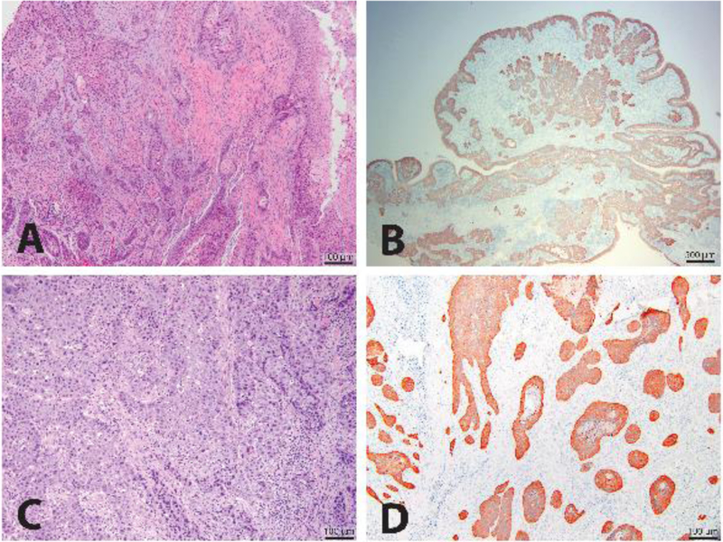
Figure 1.

Histologic subtypes of feline OSCC. A representative image of the histologic appearance of a well-differentiated conventional feline OSCC stained with H&E is shown (10X magnification) (A). A representative image for a papillary subtype of feline OSCC also stained for cytokeratins by IHC is shown (20X magnification) (B). A representative image for a basaloid subtype of feline OSCC stained with H&E is shown (100X magnification) (C). A representative image of a well-differentiated conventional feline OSCC shows all neoplastic epithelium staining positive for cytokeratins when stained with an anti-pan-cytokeratin antibody and analyzed by IHC (100X magnification) (D).
