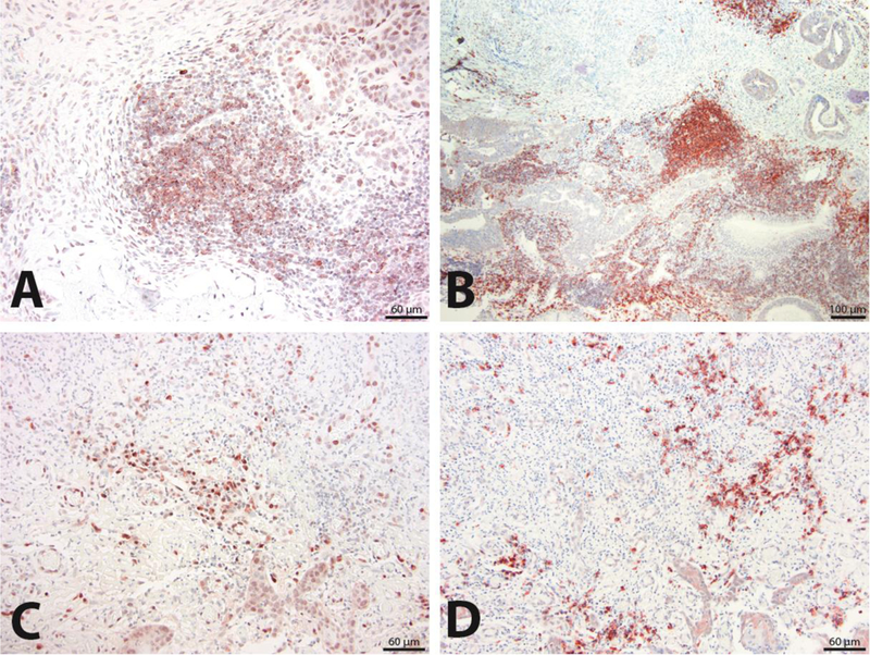
Figure 3.

Comparison of staining with CD79a and CD20 antibodies for characterization of B cell infiltrates. Tissue sections from the same feline OSCC patient biopsy analyzed by CD79a IHC (200X magnification) (A) and CD20 IHC (100X magnification) (B) are compared for detection of B cell infiltrates that also reveal follicle-like structures within neoplastic stroma. Similarly, tissue sections from a second patient biopsy analyzed with CD79a IHC (200X magnification) (C) and CD20 IHC (200X magnification) (D) are compared for detection of B cell infiltrates characterized by scattered B cells within neoplastic stroma.
