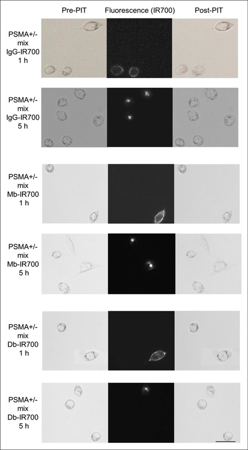
FIGURE 1.
Differential interference contrast and fluorescence microscopy of photoimmunotherapy-treated mix of PC3pip (PSMA-positive) and flu (PSMA-negative) cells. Cells were incubated with anti-PSMA antibodies (IgG-IR700), Mbs (Mb-IR700), or Dbs (Db-IR700) at 10 μg/mL. Just after fluorescence imaging, only PSMA-positive cells demonstrate necrotic cell death (cell budding/rupture). Scale bar 5 50 μm. PIT 5 photoimmunotherapy.

