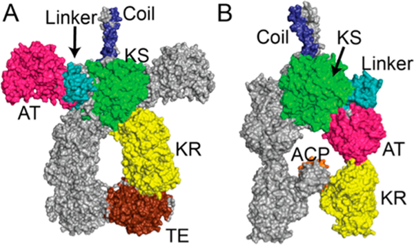
Figure 2.
Alternative conformational models of homodimeric PKS modules: (A) SAXS model of DEBS module 3 + TE;5 (B) cryo-EM model of module 5 of the pikromycin synthase.6 Docking domain (“coil”), blue; KS, green; KS-AT linker, cyan; AT, pink; KR, yellow; ACP, orange; TE, brown. The collinear domain organization is as shown in Figure 1. The two conformations differ primarily in the orientation of the AT and L domains relative to the KS.

