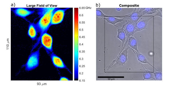
Fig. 4.
a) Example of a large field of view Brillouin shift image. The image is 110µm by 93µm, sampled at 0.5µm steps amounting to a total number of 40,920 pixels at 50ms exposure time, and total acquisition time of 34 min. Some of the cells in the image are only partly inside the horizontal confocal slice. Several nucleoli that fall within the confocal slice are visible. The lowest value of the colormap that corresponds to the measurement of cell medium is rendered black to better separate the cells from the background. b) Composite brightfield and nuclear fluorescence image of the scanned region.

