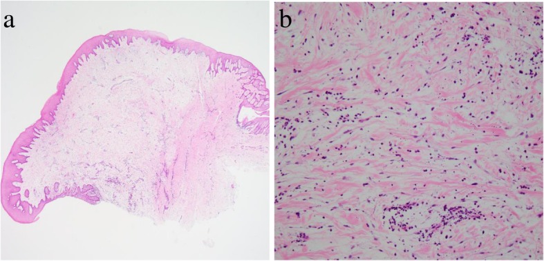
Fig. 6.

Histopathology of Case 3. a Low-power magnification (hematoxylin and eosin stain, × 20 magnification). b High-power magnification, showing myxomatous stroma with a sparsity of fibers and mild infiltration of plasma cells around the periphery of blood vessels (hematoxylin and eosin stain, × 200 magnification)
