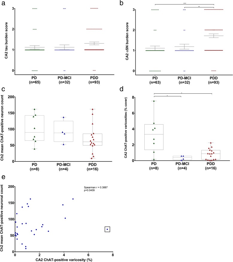
Fig. 4.
CA2 hippocampal subfield proteinaceous pathology burden and septohippocampal cholinergic deficit in PD with varying degrees of cognitive deficit. Scatter plots comparing tau (a) and αSN (b) pathology in PD, PD-MCI and PDD. Note that 7 diagnostic slides for tau analysis and 9 for αSN analysis had to be excluded due to poor tissue quality and the difficulty in accurately identifying the CA2 subfield. Error bars showing mean ± SEM. Box-and-whiskers plots comparing nvlDBB (Ch2) mean cholinergic neuron count (c) and CA2 ChAT-positive varicosities (d) between PD, PD-MCI and PDD cases. Correlation scatter graph showing the relationship between Ch2 mean cholinergic neuronal count and CA2 hippocampal subfield ChAT-positive varicosity (e). An outlier was observed and is indicated by a box. *p < 0.05; **p < 0.01; ****p < 0.0001

