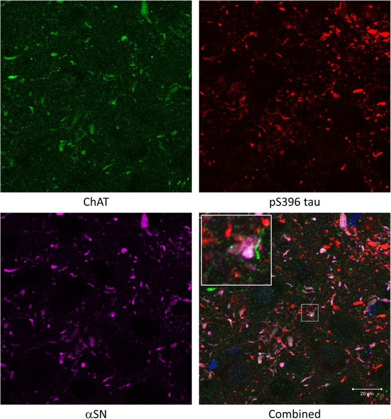
Fig. 7.

Triple immunofluorescence staining between ChAT (green), αSN (magenta) and pS396 tau (red). DAPI (blue) was also shown in combined image. A high degree of colocalisation between ChAT and αSN was observed and pS396 tau did not seem to colocalise with ChAT or αSN. Insets showing co-localisation in higher magnification. Scale bar = 20 μm
