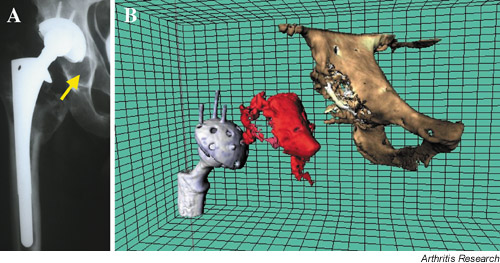
Figure 1.

Plain film and 3D segmented CT of periacetabular osteolysis. The right hip of a patient with periacetabular osteolysis was imaged by plain film X-ray (A) and computerized tomography (CT) scan. The CT scan was analyzed using artifact suppression and 3D segmentation and then reconstructed by computer (B). The virtual reconstruction shows, separately, the femoral and acetabular components of the prosthesis and the pelvic bones. The reconstruction of the region of periprosthetic osteolysis is shown between these two structures.
