
Figure 4.
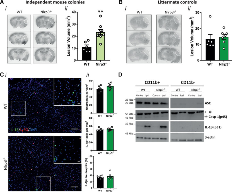
Influence of NACHT, LRR and PYD domains-containing protein 3 (NLRP3) gene deletion on brain damage after stroke. A, (i) Representative cresyl violet staining and (ii) lesion volume quantification 24 h after stroke in wild-type (WT) and nonlittermate NLRP3−/− mice (n=6–9/group; **P<0.01, analysed by 2-tailed unpaired t test). B, (i) Representative cresyl violet staining and (ii) lesion volume quantification 24 h after stroke in WT and littermate NLRP3−/− mice (n=6–7/group; ns denotes nonsignificant, analysed by 2-tailed unpaired t test). C, (i) Representative immunostaining of IL (interleukin)-1β (green), neutrophils (Ly6G, red), and 4’,6-diamidine-2’-phenylindole dihydrochloride (DAPI; blue) in the infarct 24 h after stroke (scale bar in the large image is 100 μm, and in the inset is 20 μm). C, (ii) Numbers of neutrophils and IL-1β positive cells per mm2 and % IL-1β positive neutrophils in the infarct 24 h after stroke in WT and littermate NLRP3−/− mice (n=5–6/group). D, ASC (an apoptosis-associated speck-like protein containing a CARD), Casp (caspase)-1, IL-1β, and β-actin Western blot of cell lysates from magnetic bead cell isolation of myeloid cells (CD11b+) and nonmyeloid cells (CD11b-) from the ipsi and contralateral hemisphere 24 h after stroke (n=3). *Nonspecific band.