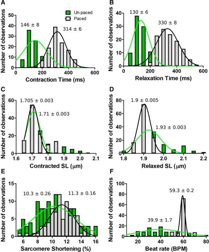
Figure 4.

Effects of electrical pacing on TTN-GFP human induced pluripotent stem cell–derived cardiomyocytes (hiPSC-CMs) contractile parameters assessed by SarcTrack. A, Histogram showing the number of observations as a function of contraction time in paced (gray, 1 Hz stimulation) and unpaced (green) hiPSC-CMs. B, Relaxation time as a function of pacing status. C, Contracted sarcomere length (SL) as a function of pacing. D, Relaxed SL as a function of pacing. E, Sarcomere shortening as a function of pacing. F, Beat rate as a function of pacing. TTN-GFP hiPSC-CMs were studied at day 30 of differentiation. Observation denotes the mean data from one field of view (≈200 sarcomeres). Histograms were generated from 122 unpaced and 116-paced fields of views. Each histogram was fitted with a single Gaussian distribution and the mean and SEM is indicated.
