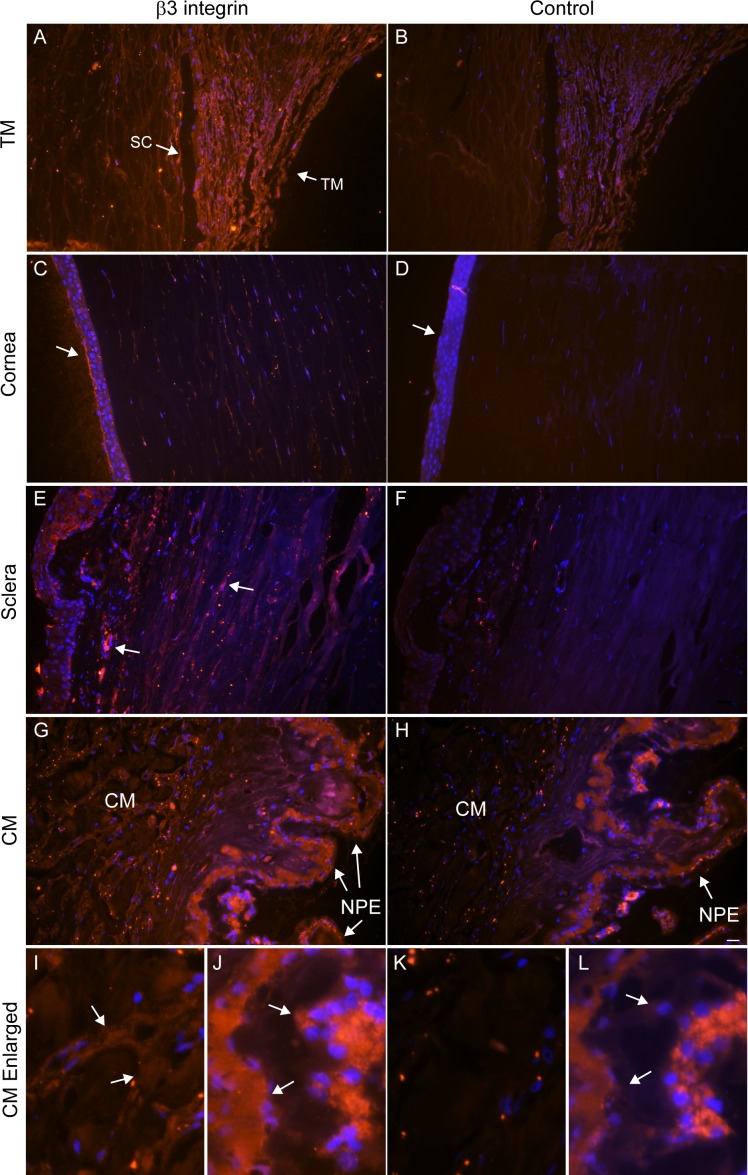
Figure 2.
Labeling of αvβ3 integrin in human anterior segments. Sections of human anterior segments from a 93-year-old donor were labeled with monoclonal antibody (BV3) against αvβ3 integrin (A, C, E, G, I, J) or a control antibody (β-galactosidase) (B, D, F, H, K, L). (A) The TM and Schlemm's canal (SC) show intense labeling for αvβ3 integrin that is clearly above the background labeling seen with the control antibody (B). (C) αvβ3 localization within the cornea. The arrow points to labeling of the corneal epithelium that is absent in the section labeled with the control antibody (D). (E) αvβ3 localization within the sclera; arrows indicate labeling absent from the section labeled with the control antibody (F). (G) αvβ3 localization within the ciliary muscle (CM) and CM nonpigmented epithelium (NPE). (H) Control antibody labeling of the same area seen in (G) that shows no labeling within the CM and NPE (arrow). (I) Enlarged view of CM shown in (G). (J) Enlarged view of NPE shown in (G). Arrows indicate positive αvβ3 labeling in these regions that is absent from the section labeled with the control antibody (K, L). Scale bar: 20 μm. Similar results were observed in anterior segments from a 36- and a 46-year-old donor (not shown).

