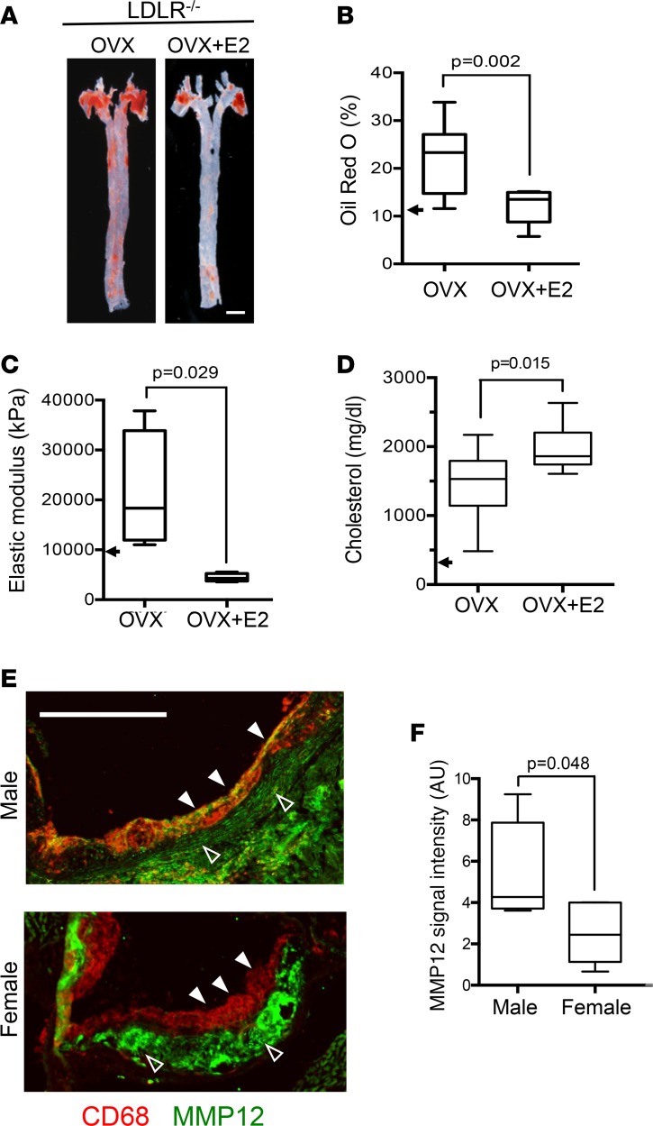
Figure 1. Protective effects of estrogen and female sex on arterial stiffness and atherosclerosis linked to reduced expression of MMP12.
(A) Representative images of atherosclerotic lesion burden of ovariectomized (OVX) LDLR–/– mice fed a high-fat diet from 8 to 24 weeks of age, with or without exogenous estradiol (OVX+E2). Lipid-rich lesions were identified by en face Oil Red O staining in aortas from OVX (n = 10) and OVX+E2 groups (n = 12). Scale bar: 1 mm. (B) Quantification of data from A expressed as a percentage of aortic area. (C) Arterial stiffness (elastic modulus) determined by AFM; n = 4 per group. The arrowheads in B and C represent the median Oil Red O staining and elastic moduli of 6-month female LDLR–/– mice on a high-fat diet without OVX (taken from Figure 2). (D) Blood cholesterol levels were measured after completion of the high-fat diet (n = 10 per condition). The arrow approximates the cholesterol level in C5BL/6 mice on a Western diet (71). (E) Aortic root sections of male and female LDLR–/– mice on a high-fat diet from 8 to 24 weeks costained for CD68 (red) and MMP12 (green). The images were merged to show colocalization; see Supplemental Figure 2 for individual images. Closed and open arrowheads show MMP12 levels in CD68+ and CD68– regions, respectively. Scale bar: 500 μm. (F) Quantification of MMP12 signal intensity in CD68+ regions from E (n = 5 per group). Graphs show box and whisker plots with Tukey’s whiskers; the horizontal lines of boxes represent the 25th percentile, the median, and the 75% percentile. Statistical significance for all panels was determined using Mann-Whitney tests.

