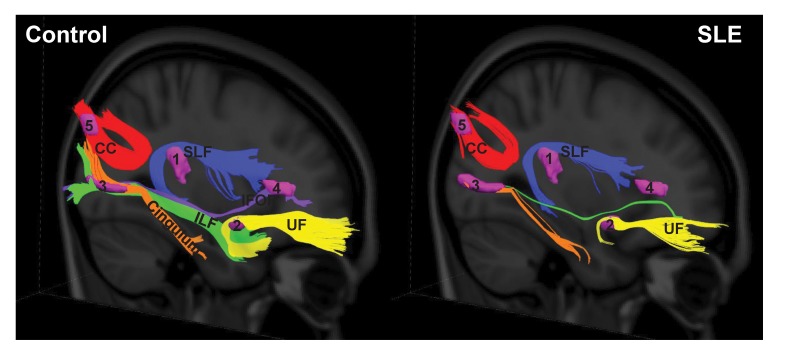
Figure 5. White matter pathways associated with the abnormal SLE-related regions visualized with group tractography.
The superior longitudinal fasciculus (temporal part) (SLF) (noted as 1), uncinate fasciculus (UF) (noted as 2), cingulum (hippocampus part) and inferior longitudinal fasciculus (ILF) (noted as 3), inferior frontal occipital fasciculus (IFOF) (noted as 4), and the splenium of the corpus callosum (CC) (noted as 5) pathways reconstructed in the healthy control (left) and SLE (right) groups. Fewer tracts were visualized in the SLE group relative to the controls in the SLF (temporal part) (−74%), UF (−86%), cingulum (hippocampus part) (−82%), ILF (–99.5%), IFOF (–100%), and splenium CC (−48%).

