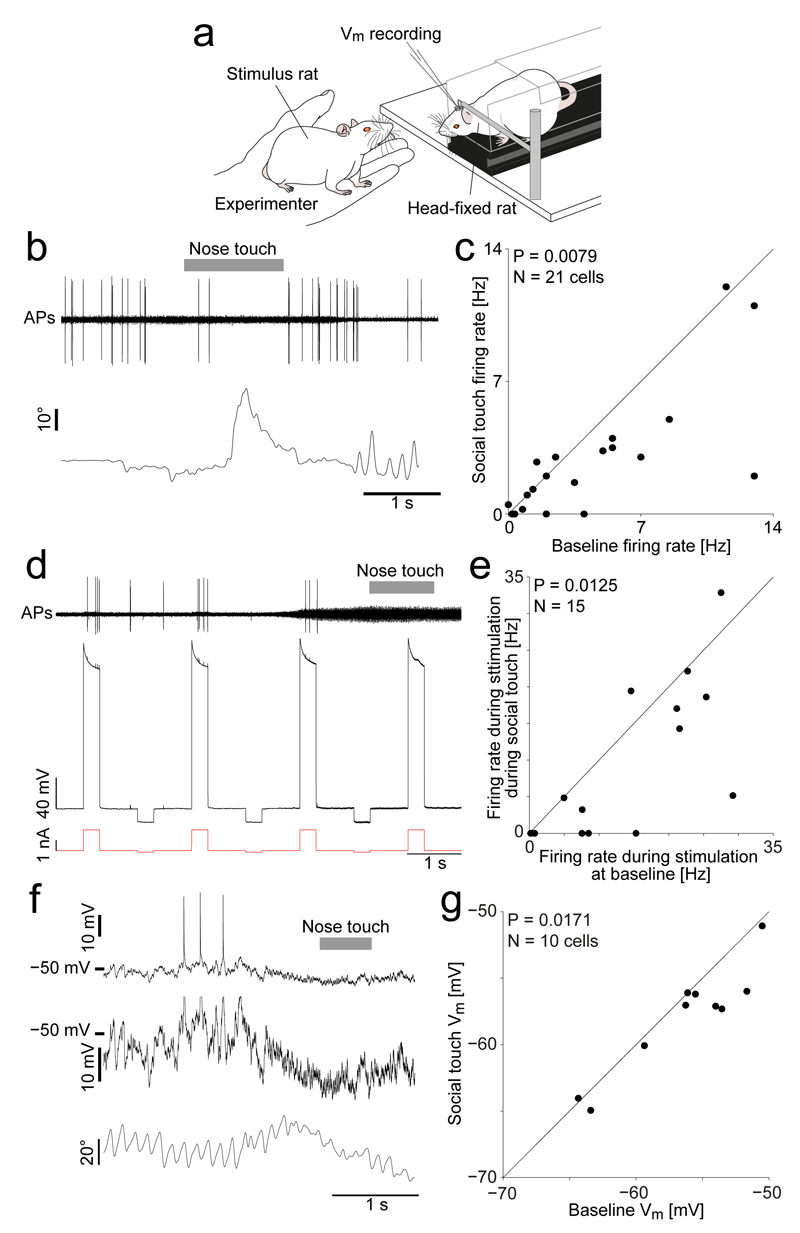
Figure 2. Decreased activity, decreased excitability and hyperpolarization of vibrissa motor cortex during social touch.
(a) VMC recording and nanostimulation in head-fixed rats during staged social touch.
(b) Top: Example juxtacellular recording in a VMC layer 5 neuron during a social facial interaction, showing a reduction in APs during social touch (nose-to-nose touch indicated by grey bar). Bottom: Angle of contralateral whisker (C2, protraction plotted upwards).
(c) Scatterplot of firing rate of VMC layer 5 cells (n=21) during social facial touch and baseline (P = 0.0079, Wilcoxon signed-rank test).
(d) Assessment of cell excitability by nanostimulation. Top: Filtered voltage trace of a VMC layer 5 cell. The evoked firing rate during nanostimulation is higher at baseline, than during social touch (indicated by grey bar). Middle: Unfiltered voltage trace. Bottom: Nanostimulation current steps.
(e) Scatterplot of the firing rate of VMC layer 5 cells (n=15) when stimulated during social facial touch and baseline (P = 0.0125, Wilcoxon signed-rank test).
(f) Top: Example whole-cell patch clamp recording from a VMC layer 5 cell showing a hyperpolarization of the membrane potential during social facial touch (duration of nose-to-nose touch indicated by grey bar). Middle: Zoom of the above trace (Spikes clipped). Bottom: Angle of contralateral whisker (C2).
(g) Scatterplot of the membrane potential (Vm) of VMC layer 5 cells (n=10) during social facial touch and baseline (P = 0.0171, t(9)=2.90, paired t-test).

