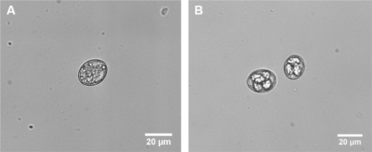
Figure 2. E. vermiformis oocysts.
This figure illustrates how “to distinguish sporulated oocyst”. A. Unsporolated or early oocyst. B. Sporulated or late oocyst, which is tetrasporic in which four sporocysts are visible. Recorded at Primovert microscope, taken at 400x magnification (Ocular 10x + objective 40x). Scale bar = 20 μm.

