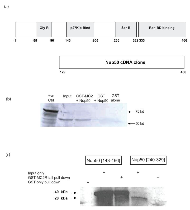
Figure 1.
GST-pull down of Nup 50. (a) depicts the domain structure of the full length Nup50 protein and the fragment of this protein encoded by the cloned cDNA identified in bacterial two-hybrid screening. (b) GST-pull down of full length Nup50 [1-466]. 35S-labelled in vitro translated Nup50 input (lane 2) was incubated with GST-MC2R tail, washed and the retained fraction separated on SDS-PAGE (lane 3). GST alone failed to pull down any 35S-labelled protein (lane 4) (c) The Nup50 [143-466] fragment is pulled down efficiently in this system (lanes 2 & 3), although little or no pull down of the smaller serine-rich domain (Nup50 [240-329]) (lanes 4 & 5) is detected.

