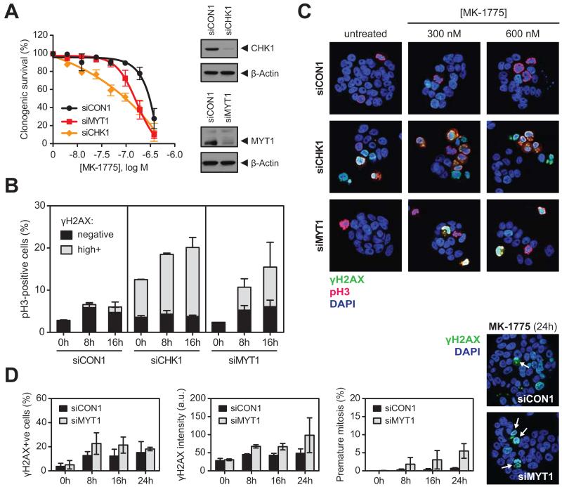
Figure 3. WEE1 inhibition promotes unscheduled mitosis after CHK1 and MYT1 depletion A.
Clonogenic survival of WiDr cells transfected with siCON1, siCHK1 or siMYT1, with colonies counted after 10 days of MK-1775 exposure. The number of colonies was normalised to untreated controls for each siRNA separately (clonogenic survival before normalisation in the absence of MK-1775: siCON1: 69%, siMYT1: 67% and siCHK1: 35%). Knockdown of CHK1 and MYT1 was confirmed by Western blot (β-Actin, loading control).
B. Flow cytometry analysis of γH2AX and pH3 after transfection with siCON1, siCHK1 and siMYT1. Cells were exposed to 200 nM MK-1775 for the indicated times. γH2AXhighpH3+ cells represent unscheduled mitosis. Error bars represent SEM of two independent experiments.
C. Immunofluorescence images showing induction of pan-nuclear γH2AX (green) and pH3 (red) staining in WiDr cells transfected with siCON1, siCHK1 or siMYT1 and exposed to 300 or 600 nM MK-1775 for 8 hours. Double γH2AX+pH3+ staining (yellow) indicates premature mitosis. DNA was counterstained with DAPI (blue).
D. WiDr cells transfected with siCON1 (black bars) or siMYT1 (grey bars) were exposed to 250 nM MK-1775 for the indicated times. Cells were stained for γH2AX and imaged by confocal microscopy. Representative immunofluorescence images show premature mitotic cells (indicated by arrows) after 24 hours of MK-1775 exposure. Bar charts show number of γH2AX-positive cells (left), γH2AX intensity (middle) and premature mitosis (right) in siMYT1-transfected cells compared to siCON1. Error bars are SEM of two independent experiments.

