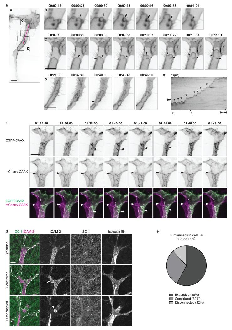
Figure 2. Apical membrane undergoes inverse blebbing during lumen expansion.
a) Embryos with mosaic expression of EGFP-CAAX were imaged from 36 hpf. Arrow in B, retracting bleb. Arrowheads in C, bleb necks. Arrow in D, lumen collapse. Time is in hours:minutes:seconds. Scale bars are 10 μm (A,C,D) and 5 μm (B). Images are representative of 7 embryos analysed.
b) A kymograph was generated along the magenta line in a, panel A. X axis, time (t) in minutes. Y axis, distance (d) in μm. Black arrowheads, retracting blebs. White arrowheads, non-retracting blebs.
c) Multicellular sprouts were imaged in Tg(kdr-l:ras-Cherry)s916 embryos with mosaic expression of EGFP-CAAX from 32 hpf. Arrowheads, inverse blebs. Time is in hours:minutes:seconds. Scale bar is 10 μm. Images are representative of 4 embryos analysed.
d) Mouse retinas were collected at P6 and stained for ICAM-2, ZO-1 and Isolectin IB4. Isolectin IB4 staining was used to draw the cell outline (white dotted line). Arrow, constricted apical membrane. Arrowhead, lumen fragment. Scale bar is 10 μm.
e) The number of lumenised unicellular sprouts showing expanded, constricted or disconnected apical membrane was quantified in P6 mouse retinas stained for ICAM-2 and ZO-1 (n=57 sprouts from 9 retinas).

