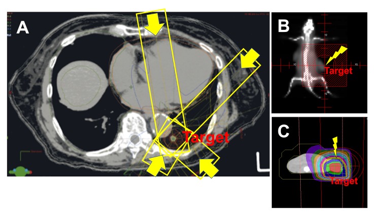
Figure 3. Difference between photon and electron beam irradiations in radiotherapies.
(A) Computed tomography (CT) of a lung cancer patient receiving stereotactic body radiotherapy (SBRT). Four representative directions of X-rays (out of 12) are marked with yellow boxes and arrows. When the photon beam (X-ray) is traversing the lung, it encounters alveoli, each of which is surrounded by a capillary network so extensive that it forms an almost continuous sheet of surrounding blood. Therefore, regardless whether lung or other tissues, harmful reactive oxygen species (ROS) seem to be produced in the peripheral blood, which are considered to reduce lymphocyte number directly or indirectly. (B) Field of radiation and (C) CT simulation of the irradiated field in a mouse model used in our preclinical studies. Tumor cells were implanted subcutaneously in both flanks. Electron beams were derivers to the right flank from just above the tumor. The beams are only effective for subcutaneously growing tumor and scarcely affect other tissues. In contrast to X-rays, electron beams are known to have a finite range, after which dose falls off rapidly, sparing underling healthy tissue with extensive blood vessel networks.

