Figure 1.
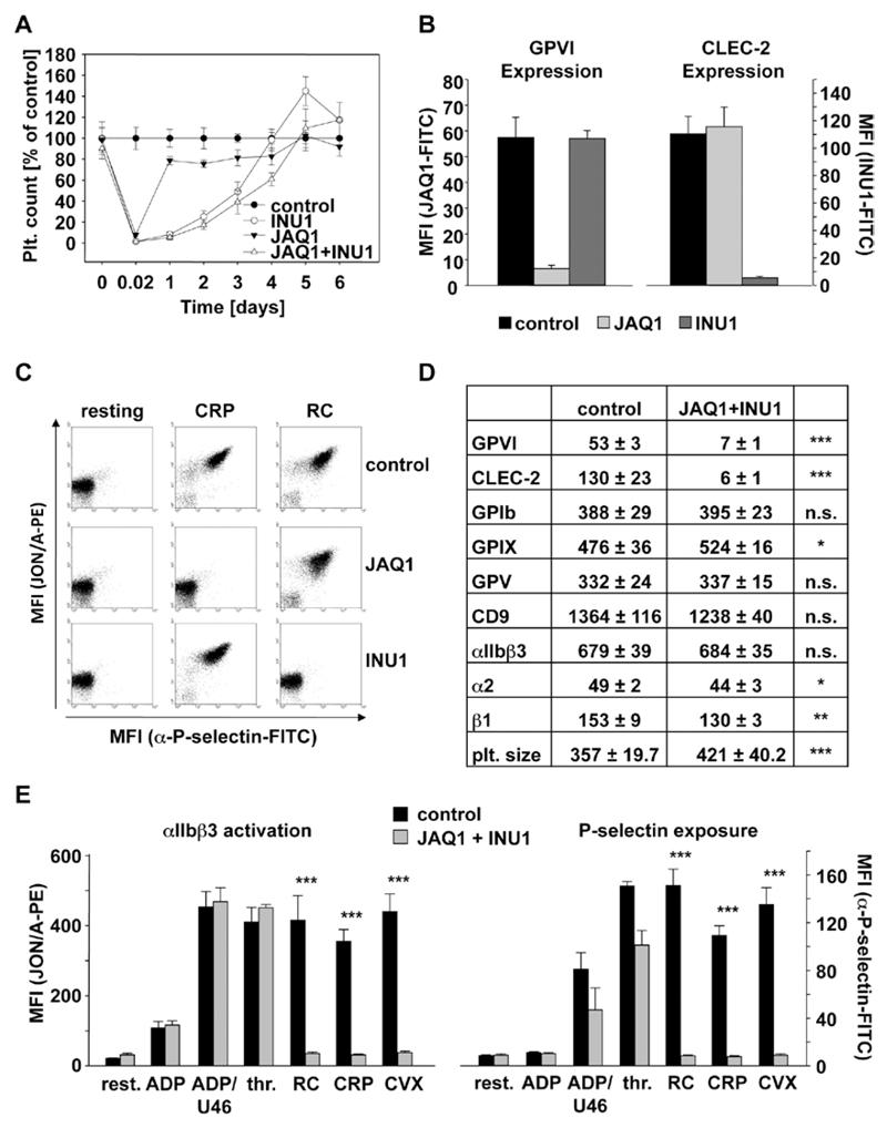
Analysis of mice deficient in Glycoprotein VI (GPVI) and C-type lectin-like receptor 2 (CLEC-2) on antibody injection. A, Mice were intravenously injected with 100 µg JAQ1 and 200 µg INU1 in sterile PBS, and platelet counts were determined on a FACSCalibur at the indicated time points post injection. Results are mean±SD in % of control animals (n=5 mice per group, representative for 2 individual experiments). B and D, Flow cytometric analysis of surface protein expression 5 days post injection with the indicated antibodies. Platelets were stained for 15 minutes at room temperature with the indicated fluorophore-labeled antibodies and directly analyzed. Platelet count in number of platelets/µL. Platelet size is given as mean forward scatter (FSC) and was determined by FSC characteristics. Results are mean fluorescence intensities (MFI)±SD (n=5, representative of at least 3 independent measurements). *P<0.05; **P<0.01; ***P<0.001. C and E, Flow cytometric analysis of integrin αIIbβ3 activation (JON/A-PE) and degranulation-dependent P-selectin exposure on platelets on day 5 post injection. Washed blood was incubated with the indicated agonists for 15 minutes and analyzed on a FACSCalibur. Results are mean±SD (n=5 mice per group, representative of 3 independent experiments). ***P<0.001. ADP: 10 µmol/L; U46619: 3 µmol/L; thrombin: 0.01 U/mL; rhodocytin (RC): 1 µg/mL; collagen-related peptide (CRP): 10 µg/mL; convulxin (CVX): 1 µg/mL. All experiments were performed on day 5 to 6 after antibody injection. FITC indicates fluorescein isothiocyanate.

