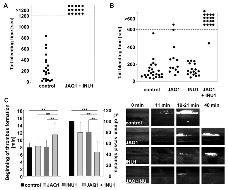
Figure 2.
Determination of hemostatic function and pathological thrombus formation in Glycoprotein VI (GPVI)/C-type lectin-like receptor 2 (CLEC-2)–depleted mice. A, A 1-mm segment of the tail tip was cut, and bleeding was determined to have ceased when no blood drop was observed on the filter paper. Each symbol represents 1 individual. B, A 1-mm segment of the tail tip was cut, and the tail tip was immersed in saline. Each symbol represents 1 individual. Differences of bleeding times between wild-type (WT), single GPVI-depleted, and single CLEC-2–depleted mice are nonsignificant. C, Mesenteric arterioles were treated with 20% FeCl3, and adhesion and thrombus formation of fluorescently labeled platelets were monitored by in vivo fluorescence microscopy. Statistical evaluation of the time to appearance of a first thrombus (left) and percentage of maximal vessel stenosis (right) are depicted, n≥10. At most 2 arterioles of each mouse were analyzed. **P<0.01; ***P<0.001. Vessel stenosis was determined by measuring maximal thrombus size divided by vessel diameter using the Metamorph software (Visitron). Representative images are shown. White asterisk indicates occluded vessel. All experiments were performed on day 5 to 6 after antibody injection.

