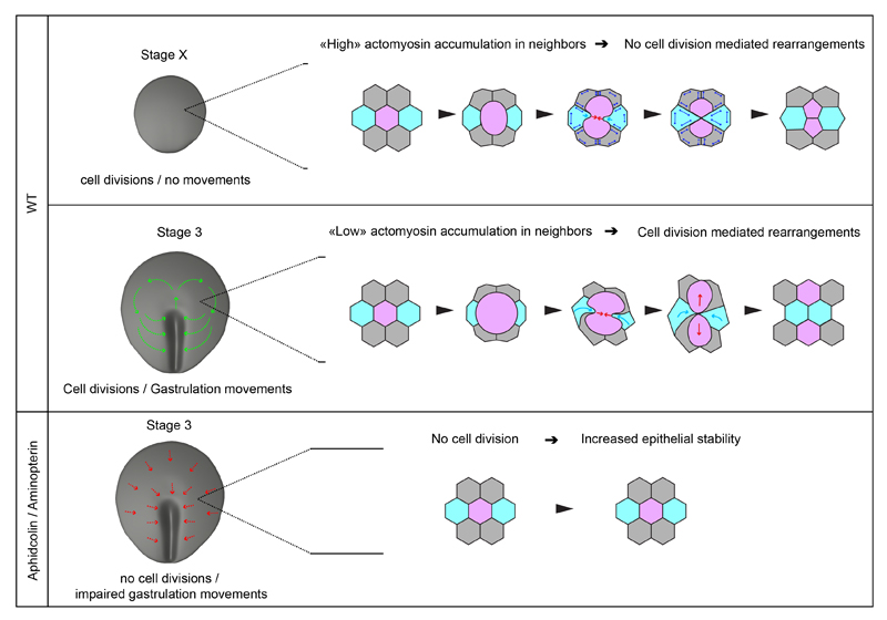
Figure 7. Model for the role and control of cell division during gastrulation.
Higher panel: at stage X, before gastrulation movements initiate epithelial cells divide without promoting rearrangements; epithelial cells exhibit higher actomyosin accumuluation: the dividing cell induces very local deformation of neighbors; as they resist deformation and exhibit high junctional stability (double arrows in blue), these cells consequently fail to move in between daughters and intercalation does not take place. Middle Panel: at stage 3, as gastrulation movements are taking place, cell division promotes epithelial cell rearrangements; epithelial cells exhibit lower actomyosin accumulation enabling dividing cells to deform and displace neighbors (light blue arrow), bringing them in between the resulting daughters. Lower panel: In absence of cell division, epithelial stability is increased as cell division mediated rearrangements do not take place; epithelial cells are likely pulled (red arrows) towards the primitive streak, where intercalation/ingression events take place (Rozbicki et al., 2015; Voiculescu et al., 2007, 2007).

