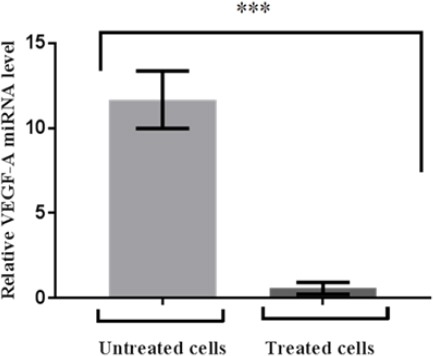
Figure 3.

The VEGF-A Expression at 24 hr of MCF7 Cells. Cells were transfected with miR-126 lipofectamine. Relative miRNA VEGF-A (normalised to GAPDH) was detected by real time RT PCR. Data are mean ± standard error of mean (n=3) and were analysed by student’s t-test (one tailed). Significantly down regulated VEGF-A levels in treated breast cancer are shown relative to VEGF-A levels in adjacent untreated (control) cells
