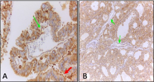
Figure 2.

Immunohistochemical Detection of VEGFR-1 in Cytoplasm and Membrane (Red Arrow) of Papillary Thyroid Carcinoma Cells (A) and cytoplasm of thyrocytes of nodular hyperplasia (B). Green arrows indicate VEGFR-1 staining in endothelial lining of blood vessels. Original magnifications, A, x 400 and B, x 100.
