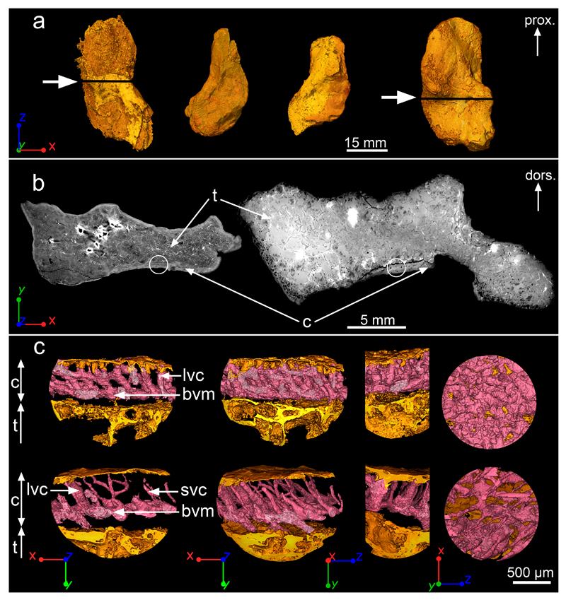
Figure 1. Midshaft bone microanatomy and histology of Acanthostega’s humerus.
a, 3D models of humeri shown in ventral view, based on synchrotron data. From left to right: NHMD 74756 (voxel size: 14.95μm), UMZC T.1295 (voxel size: 12.62μm), MGUH 29019 (voxel size: 12.62μm) and MGUH 29020 (bottom, voxel size: 20.24μm). The white arrows indicate the location of the virtual thin sections in b. b, Humeral microanatomy of NHMD 74756 (left) and MGUH 29020 (right) showing an extended trabecular cavity (t) surrounded by a thin layer of compact cortical bone (c). The transverse virtual thin sections are 80μm thick. The white circles indicate the location of the high-resolution scans modelled in c. c, 3D models (voxel size: 0.638μm) of the cortical bone microstructure of NHMD 74756 (top) and MGUH 29020 (bottom) showing a dense oblique mesh of large (lvc) and small (svc) vascular canals (in pink) connected to a horizontal basal vascular mesh (bvm). From left to right: transverse 3D thin section (250μm thick), complete 3D model in transverse orientation, 3D model in longitudinal orientation, tangential section showing the inner view of the 3D vascular mesh. Abbreviations: dors., dorsal; prox., proximal.

