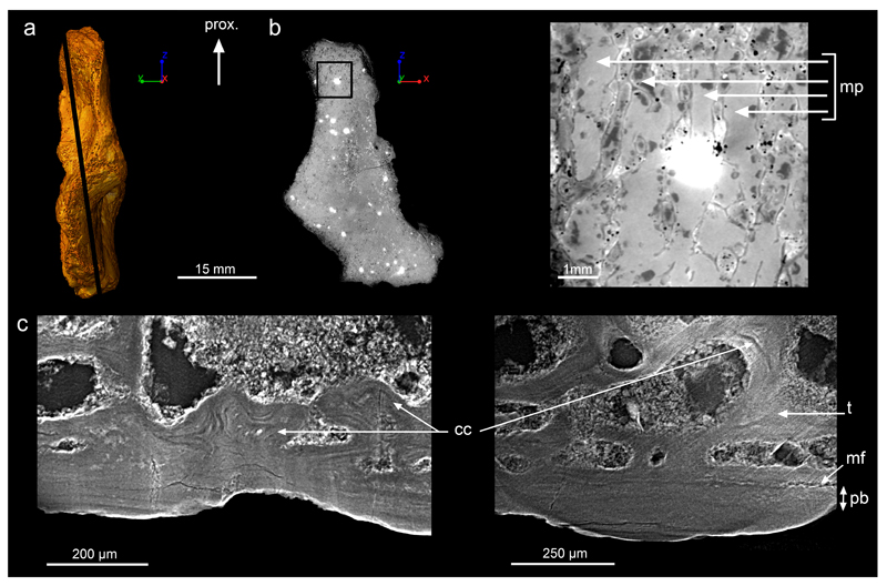
Extended Data Figure 2. Epiphyseal microanatomy and histology of Acanthostega’s humerus (MGUH 29020).
a, 3D model in preaxial view, based on synchrotron microtomography data. It is oriented with the proximal extremity (epiphysis6) towards the top. The black line indicates the virtual thin section illustrated in b. b, Longitudinal virtual thin section (thickness: 50μm, voxel size: 1.12μm, same scale bar and orientation as in a) showing the location where the detailed image on the right comes from. The latter shows the marrow processes (mp) formed in the growth plate by endochondral ossification. c, High-resolution virtual thin section (thickness: 50μm, voxel size: 1.12μm) from the epiphyseal region showing obvious Liesegang’s rings as remnants of calcified cartilage6 (cc), formed during endochondral ossification. These remnants are entrapped in the trabeculae (t), at the vicinity of the ossification notch6, where the thickness of the periosteal bone (pb) – between the mineralisation front (mf) and the surface – drastically reduces. The bone is oriented with its surface towards the bottom. From left to right: longitudinal thin section, transverse section. Abbreviation: prox., proximal.

