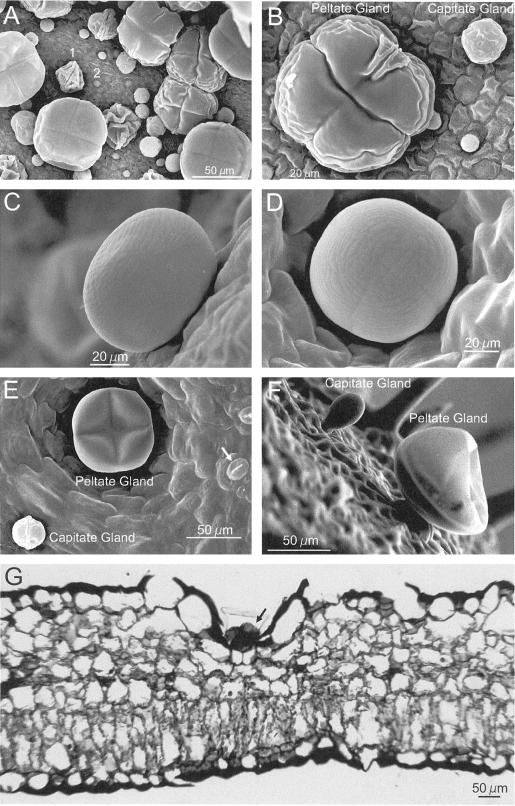
Figure 2.
Morphology of basil peltate and capitate glands. A, SEM of adaxial surface of a very young EMX-1 leaf (<0.5 cm long) showing the high density at initiation of peltate and capitate glands on basil leaves. 1, Peltate gland early in development; 2, gland primordium. B, Closer view of peltate and capitate glands from same leaf as in A. The 4- and 2-fold symmetries, respectively, of each gland type are clearly visible, as are two perpendicular creases and the wrinkled folds of the immature oil sac membrane. C, Side view of a fully expanded peltate gland on a young EMX-1 leaf (1.5 cm long). D, Peltate gland on the surface of a young EMX-1 leaf, showing the fine structure of the oil sac, including the two perpendicular remnant creases left by the four underlying secretory cells. E, Adaxial surface of an expanding EMX-1 leaf (2 cm long). The peltate gland, whose oil sac is partially deflated, is recessed into the surface of the leaf. Stomatal guard cells are visible (arrow). F, Structure of glands on the surface of developing sepals from line SW, revealing the stalk cell connecting each gland to the leaf. Several non-glandular trichomes can be seen in the background. Again, the oil sac on the peltate gland is partially deflated. G, Light micrograph of an EMX-1 leaf cross-section (adaxial surface up) stained with toluidine blue showing how deeply the peltate glands (arrow) are recessed into the leaf.

