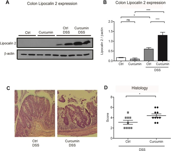
Fig 3. Curcumin supplementation in DSS-treated mice enhances inflammation in the colon.
(A) Representative western blot of colon protein extracts probed with antibodies against lipocalin 2 and β-actin. Each lane represents an individual mouse. (B) Graphic depicting densitometric quantification of western blots from three independent experiments. Data are presented as mean ± SEM. (C) Representative hematoxylin and eosin staining of mouse colon histological sections. (D) Graphic depicting quantification of colonic histology scores. Statistical analysis was performed by two-tailed Student’s t-test. *P < 0.05, **P < 0.01, ***P < 0.001; ns = not significant; n = 8 mice per group.

