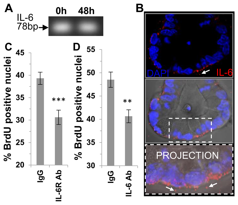
Figure 3. Autocrine IL-6 signaling regulates small intestinal organoid proliferation.
(A) PCR gel showing IL-6 transcript expression in in vivo / freshly isolated (0 h) and cultured (48 h) small intestinal crypts (B) Representative confocal image showing immunofluorescent labelling of IL-6 (red), nuclei (DAPI blue) of small intestinal organoids with the DIC image overlayed and associated projection image in vitro. White arrows indicate extracellular pools of IL-6. Scale bar 10 μm. (C) Histogram showing percentage positive BrdU positive nuclei in mouse small intestinal organoids in the presence of an IL-6 receptor blocking antibody (n=3, **P<0.01) and (D) an IL-6 neutralising antibody (n=3, ***P<0.001) compared to respective IgG control. Data are represented as mean +/- SEM.

