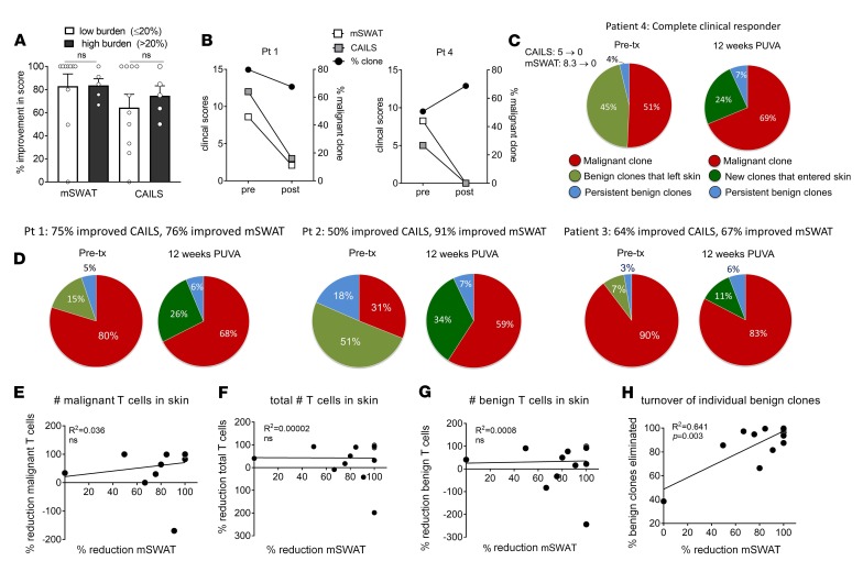
Figure 2. Visible inflammation does not reflect malignant T cell burden and reduced inflammation is linked to turnover of benign T cells.
(A) Clinical exam scores in high- and low-burden patients were not significantly different. (B) Two patients are shown in whom visible inflammation (clinical exam scores) improved but the malignant T cell clone remained high after treatment (patient 1, 68%; patient 4, 69%) or even increased (patient 4). (C) Malignant T cell frequency remained high after treatment despite the presence of large numbers of malignant T cells in skin in patient 4, a complete clinical responder. The unique TCR CDR3 sequences of each nonmalignant T cell clone were used to identify which benign T cells persisted after therapy (blue), were eliminated from skin (light green), or were recruited to skin (dark green) after therapy. Persistent benign clones were benign T cell clones that were present in skin both before and after PUVA therapy. (D) Additional patients are shown in whom the malignant T cell burden remained high after therapy despite improvement in clinical inflammation exam scores. (E–H) Improvement in inflammation is correlated with a shift in the benign T cell population but not with depletion of malignant T cells. Improvement in inflammation (mSWAT) did not correlate with reductions in the number of malignant T cells (E), total T cells (F), or benign T cells (G). However, the loss of specific T cell clones from skin and recruitment of a second, distinct T cell population was correlated with reduced inflammation as assessed by mSWAT (H) and CAILS scores (Supplemental Figure 1). Differences between 2 sample groups were detected using the 1-tailed Wilcoxon-Mann-Whitney test (α = 0.05). For correlations, a Pearson’s correlation coefficient with a 2-tailed P value is reported.

